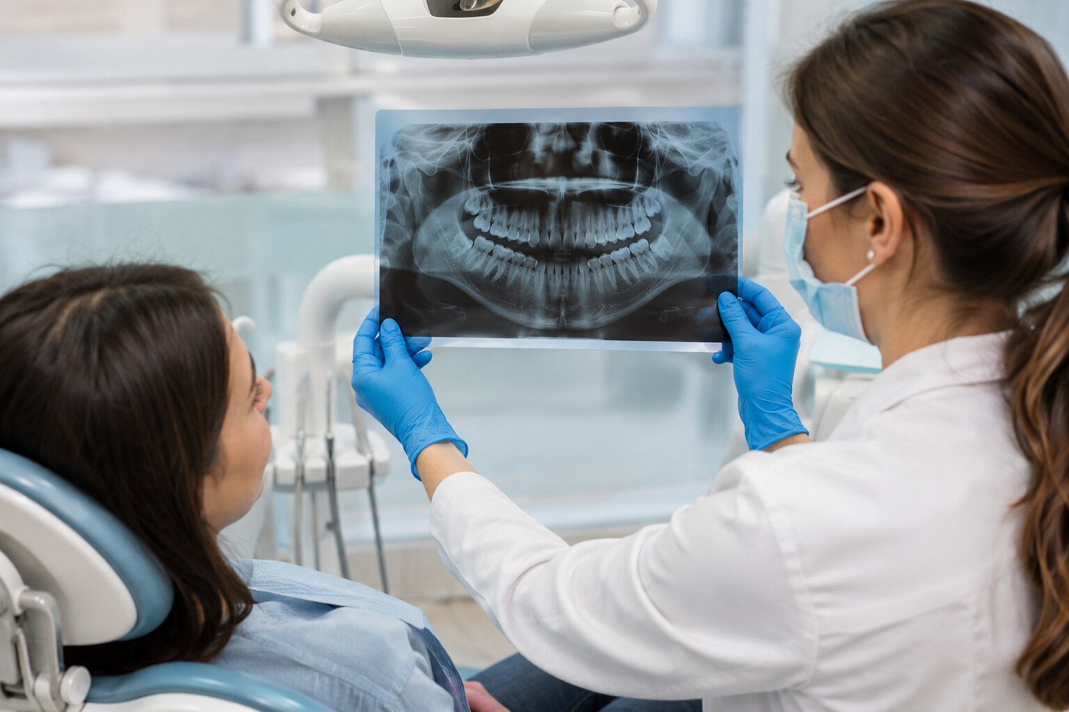
A tooth can look intact while decay between teeth, root infection, bone loss, an impacted tooth or a fracture remains hidden from view. By the end, you will know which symptoms and treatment decisions justify an X-ray, which view answers each question, and when imaging adds little useful information.
Key takeaways
- Ask why the image is needed and what treatment decision it will change.
- A bitewing X-ray detects between-tooth decay; a panoramic view surveys jaws and developing teeth.
- Standard X-rays miss some soft-tissue problems and fine three-dimensional details.
- CBCT is reserved for complex cases such as impacted teeth, implants, and surgical planning.
When does a dentist need an X-ray for a hidden tooth problem?
When is a dental X-ray needed? When examination findings suggest a hidden tooth problem that could change treatment. The dentist considers your symptoms, dental history, age, previous images, decay or gum-disease risk, and what the image would clarify.
1. During preventive care, a routine dental X-ray may be a bitewing image if you have higher decay risk or suspected decay between teeth. Children and adolescents do not automatically need a full-mouth series; age, developing teeth, exposure history and clinical findings guide the decision.
2. A symptom-driven dental X-ray matches the complaint. Pain in one tooth without a visible cavity may need a targeted periapical image showing that tooth, its roots and surrounding bone. The same view can investigate a dental abscess, unexplained swelling, trauma, failed root-canal symptoms or deep decay.
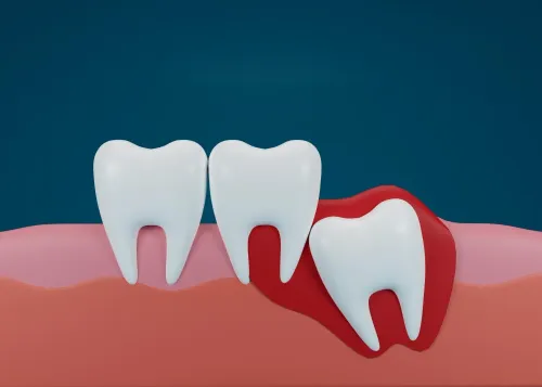
3. Imaging can also support assessment of loose teeth or bone loss, impacted or erupting wisdom teeth, and planning before extraction, root-canal treatment, orthodontics or another procedure. A panoramic image may suit a broad jaw question, but it is not automatically useful for every complaint.
Dentists avoid ordering the same view for every patient because a full-mouth series adds exposure and information that may not affect care.
Ask, “What clinical question will this X-ray answer?” and “Will the result change treatment?” Tell the dental team if you are pregnant or could be pregnant, so timing and technique can be considered appropriately.
Which dental X-ray view reveals each type of hidden problem?
Match the suspected hidden problem to the view that captures it, rather than taking one broad image for every complaint.
| View | What it shows | Most useful for |
|---|---|---|
| Bitewing X-ray | Crowns of upper and lower back teeth together, plus the bone level between them | Decay between teeth and assessment of periodontal bone loss |
| Periapical X-ray | One tooth from crown through root tip, with surrounding bone | Suspected abscess, trauma, deep decay, root development and endodontic assessment |
| Panoramic X-ray | Both jaws, the temporomandibular-joint region, and developing or unerupted teeth | Impacted wisdom teeth, orthodontic assessment, extensive jaw disease and some surgical planning |
| Occlusal X-ray | A larger section of the mouth using a sensor placed across the biting surface | A wider area when a standard intraoral image does not cover enough tissue |
A panoramic X-ray provides useful orientation but less fine detail than an intraoral image, so it is not the best test for a small cavity between two teeth. A bitewing X-ray answers that question more directly.
For pain localised to one tooth, a periapical X-ray may provide the needed evidence without a full-mouth series. Occlusal imaging becomes useful when swelling, an unerupted tooth or another finding extends beyond the area captured by a standard sensor. The clinical question determines the view.
What can a dental X-ray show, and what can it miss?
Dental X-rays show mineralised decay, tooth roots, impacted teeth, changes in surrounding bone, bone loss and some infections. They are most useful when another tooth, a restoration, bone or an adjacent tooth surface hides the area during direct inspection.
A bitewing can reveal decay between back teeth, while a localised periapical image shows the full tooth and root-area bone.
A normal image does not clear every cause of pain. Early decay, a tooth crack X-ray when the crack does not open in the beam, pulp inflammation and soft-tissue conditions can remain unclear or invisible. These dental X-ray limitations mean the image supports examination rather than replacing it.
The dentist may combine the image with:
- Visual inspection
- Tapping and bite testing
- Periodontal measurements
- Pulp-vitality testing
A painful tooth does not automatically justify a panoramic image or full-mouth series. If symptoms and examination localise the problem, a periapical view may answer the question with less exposure and less incidental information.
The dentist can defer imaging when its result would not change diagnosis or treatment; ordering it makes sense when a hidden finding could alter care.
When is 3D CBCT needed instead of a standard dental X-ray?
Choose a CBCT dental scan when a standard two-dimensional image and examination leave a specific treatment question unanswered. Cone-beam computed tomography provides 3D dental imaging that can reveal the three-dimensional position of an impacted tooth, complex root anatomy, selected difficult endodontic problems, implant-planning anatomy, and some jaw lesions or oral-surgical problems.
It is not a routine replacement for bitewing, periapical, panoramic, or occlusal radiographs. A painful tooth does not automatically need CBCT; a targeted periapical image may answer the question with less exposure.
CBCT generally delivers more radiation than a conventional dental radiograph. Dose varies with the machine, field of view, and scan settings, so request the smallest field of view and lowest exposure that can answer the clinical question.
An implant planning X-ray may be insufficient when the dentist must measure bone width or locate nearby structures in three dimensions, but advanced imaging is not automatically more accurate or necessary.
Ask these questions before agreeing to the scan:
- What information is missing from the 2D image?
- Will the 3D result change the procedure, such as implant placement, removal of a difficult impacted tooth, or complex root treatment?
- Can a limited field of view answer the question?
If the answer to the second question is no, CBCT may add radiation and incidental findings without improving your care.
What happens during dental imaging, and how can you limit unnecessary exposure?
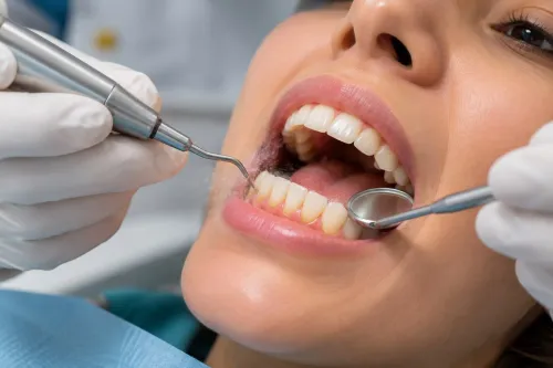
An intraoral dental X-ray procedure involves biting gently on a holder or digital dental sensor while the team positions it against your cheek or palate. The holder can feel awkward, but it should not hurt.
A panoramic image requires you to stand or sit still with your chin and head aligned in the machine; the exposure itself takes only a short time.
Movement, incorrect positioning or metal objects in the field can blur or obscure the image and lead to an X-ray retake. Follow the bite instructions, keep still and remove glasses, earrings or removable metal appliances if asked.
Use this dental X-ray radiation safety checklist:
- Tell the dentist if you are pregnant or could be pregnant. Pregnancy alone does not rule out a necessary image; disclosure lets the team consider timing and exposure settings.
- Share recent images from another clinic, including the date and view taken, so the team can avoid needless repeats.
- Ask whether a digital sensor is being used and whether the clinic follows current local radiation-protection guidance.
- Ask which finding the image will investigate and how the result could change treatment. At PRYSM DENTAL STUDIO, use these questions when discussing targeted imaging or pre-treatment planning.
| Image choice | Positioning | Main reason to avoid a broader study |
|---|---|---|
| Intraoral | Bite on a holder or sensor | A focused question needs only a small area |
| Panoramic | Align chin and head in the machine | A broad view may add detail that does not affect care |
| CBCT | Keep still inside the scanner | Use the smallest field and lowest exposure that answers the question |
Related service
Digital Dental X-Rays in Deccan Gymkhana, Pune – Fast, Safe & Accurate Diagnosis At Dr. View service → |
Frequently asked questions
When does a dentist need an X-ray for a hidden tooth problem?
An X-ray is needed when examination findings, symptoms, dental history, age, or disease risk suggest a hidden problem that could change treatment. Ask what the image will clarify before having it taken.
Which dental X-ray view reveals each type of hidden problem?
Bitewing X-rays show decay between teeth and bone levels near the crowns. Periapical X-rays show the full tooth, roots, and surrounding bone. Panoramic X-rays survey both jaws, impacted teeth, and developing teeth. Occlusal views show a larger section of one arch.
What can a dental X-ray show, and what can it miss?
Dental X-rays can show cavities, bone loss, root infections, fractures, impacted teeth, and changes around roots. They do not show every soft-tissue problem and can miss early decay, fine cracks, or details hidden by overlapping structures.
When is 3D CBCT needed instead of a standard dental X-ray?
CBCT is used when three-dimensional information will affect care, such as locating an impacted tooth, mapping roots near a nerve, planning implants, or assessing complex surgery. A standard X-ray is preferred when it answers the clinical question.
What happens during dental imaging, and how can you limit unnecessary exposure?
You will hold a sensor or imaging plate in position while the machine takes a brief image; CBCT involves remaining still while the scanner rotates around your head. Share previous images, ask whether a new image will change treatment, and use the smallest suitable scan.