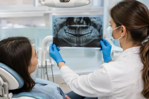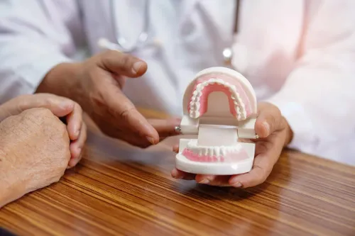
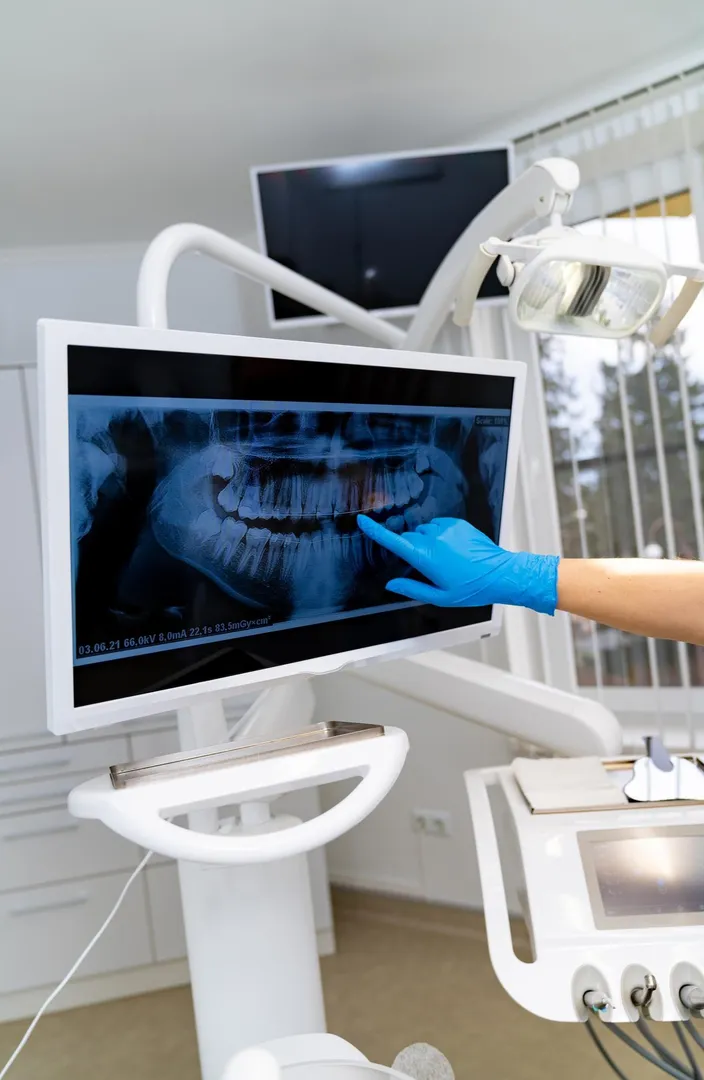
Dental problems are not always visible during a routine examination. Decay can develop between teeth, infections may form around tooth roots, and changes in the supporting bone can occur without obvious symptoms. Digital Dental X-Rays help dentists examine these hidden areas and make more informed decisions about further evaluation and treatment.
For patients in Pune and across Maharashtra, digital imaging has become an important part of modern preventive and restorative dental care.
What Digital Dental X-Rays Can Show
A visual dental examination provides valuable information about the teeth and gums, but it cannot reveal everything happening below the surface. Depending on the type of image taken, dental X-rays may help identify:
- Decay between teeth
- Changes around tooth roots
- Bone levels supporting the teeth
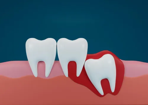
- Impacted wisdom teeth
- Developing permanent teeth in children
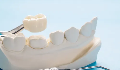
- Existing fillings, crowns, and other restorations
- Areas that may require further investigation
The dentist determines whether an X-ray is necessary based on symptoms, dental history, age, examination findings, and the treatment being considered.
How Digital Imaging Differs from Traditional Dental X-Rays
Digital dental radiography uses electronic sensors or digital imaging systems rather than conventional film.
The resulting images can usually be viewed quickly on a computer screen. Dentists can enlarge particular areas and examine structures in greater detail when required.
Digital storage can also make it easier to compare current images with previous records. This can be useful when monitoring changes in a tooth, bone levels, restorations, or developing dental conditions over time.
When Your Dentist May Recommend an X-Ray
Not every appointment requires an X-ray. Imaging is generally recommended when the expected diagnostic benefit justifies it.
A dentist may consider imaging when a patient has persistent tooth pain, sensitivity, swelling, suspected decay, trauma, unexplained changes around a tooth, or symptoms associated with wisdom teeth.
X-rays may also support planning for procedures such as root canal treatment, dental implants, orthodontic care, extractions, crowns, and other restorative treatments.
Children may sometimes need dental imaging to evaluate tooth development and eruption patterns. The need and frequency should be decided individually.
Why Early Detection Matters
Some dental conditions are easier to manage when identified before they become extensive.
For example, decay detected at an earlier stage may require a different treatment approach than a tooth that has developed deep decay involving the pulp. Similarly, identifying changes in the supporting bone can help the dentist investigate possible periodontal concerns.
However, an X-ray is only one part of diagnosis. Findings should be interpreted alongside the patient’s symptoms, clinical examination, dental history, and other relevant information.
What Patients Can Expect
The imaging process is generally straightforward. Depending on the area being examined, a small sensor may be positioned inside the mouth while the X-ray image is taken. Other imaging systems can capture a wider view of the jaws and teeth.
Patients should inform the dental team about pregnancy or any other information that may be relevant before imaging.
At PRYSM DENTAL STUDIO, digital imaging can be incorporated into a broader dental assessment when clinically appropriate. For Pune patients, this means treatment planning can be based on information gathered from both a physical examination and relevant diagnostic images.
Digital Dental X-Rays are therefore best viewed as a diagnostic aid rather than a treatment themselves. When used appropriately, they give dentists another way to understand what is happening beneath the visible surface and determine whether monitoring, preventive care, or treatment should be considered.
Keywords